
When Diagnostic Imaging for Back Pain Helps
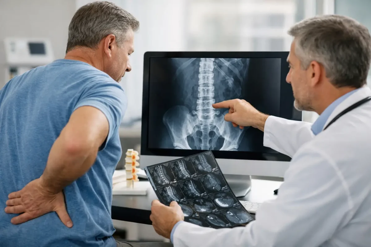
A painful back can make every movement feel like a question: Is this a muscle strain, a disc problem, arthritis, or something more serious? Diagnostic imaging for back pain can provide useful answers, but the right scan at the right time matters more than getting imaging as quickly as possible. For many people, a careful examination and conservative treatment are the appropriate first steps. For others, imaging can help identify a condition that needs more targeted care.
At Denville Medical Associates, back pain evaluations are designed to connect the clinical picture with a practical recovery plan. That means considering your symptoms, medical history, physical examination, activity demands, and imaging results together rather than treating a scan as the whole answer.
Why a Scan Is Not Always the First Step
Back pain is common, and many episodes improve within several weeks with appropriate non-surgical care. Muscle and ligament strains, minor joint irritation, and uncomplicated flare-ups of chronic low back pain often do not require immediate imaging. In these cases, early X-rays, CT scans, or MRIs may reveal age-related changes that are not actually causing the pain.
For example, disc bulges, spinal arthritis, and mild degeneration are common findings in adults who have no back pain at all. Seeing those changes on a report can create understandable worry, yet it does not automatically mean they need injections or surgery. A meaningful diagnosis comes from matching what appears on an image with where your symptoms occur, how they behave, and what is found during a physical exam.
This approach can also reduce unnecessary radiation exposure, avoid incidental findings, and help patients begin treatment without waiting for a test that may not change the initial plan. Physical therapy, guided activity modification, anti-inflammatory strategies when appropriate, chiropractic care, acupuncture, or pain management may be recommended based on the individual situation.
When Diagnostic Imaging for Back Pain Is Recommended
Imaging becomes more valuable when it is likely to change the course of care. A physician may recommend it after a significant fall, sports injury, work injury, or motor vehicle accident, especially when there is concern for a fracture or structural injury. It may also be appropriate when pain has not improved after a reasonable period of conservative treatment.
Certain symptoms need prompt medical evaluation because they may point to nerve compression, infection, fracture, or another condition requiring urgent attention. These include new or worsening leg weakness, numbness in the groin or saddle area, loss of bowel or bladder control, fever with severe back pain, unexplained weight loss, a history of cancer, or pain that is severe and unrelenting at night.
If back pain follows major trauma, occurs alongside chest pain or abdominal pain, or is accompanied by sudden loss of strength or bowel and bladder control, seek emergency care. These symptoms should not wait for a routine outpatient appointment.
For non-emergency concerns, imaging may help when pain travels down the leg, numbness or tingling persists, reflexes or strength are changing, or a specialist is considering an injection or surgical consultation. The goal is not simply to find an abnormality. It is to obtain information that supports a safer, more precise treatment decision.
The Main Types of Back Imaging
Different imaging studies show different structures. Choosing the test depends on the suspected cause of pain, the part of the spine involved, prior treatment, and your health history.
X-rays
X-rays are often used to evaluate bones and spinal alignment. They can help identify fractures, significant arthritis, curvature changes, vertebral slippage, and instability that may appear when the spine moves. They are quick and widely available, but they do not show discs, nerves, muscles, or other soft tissues in detail.
X-rays may be particularly helpful after an injury, when osteoporosis raises fracture risk, or when a clinician needs to assess alignment. They use a small amount of radiation, so the benefits should be weighed against the clinical need.
MRI
Magnetic resonance imaging, or MRI, provides detailed pictures of soft tissues. It is often the preferred study when a physician suspects a herniated disc, spinal stenosis, nerve root compression, infection, inflammation, or certain tumors. An MRI does not use ionizing radiation.
MRI can be highly informative for sciatica, especially when leg pain, weakness, or numbness corresponds to a particular nerve pathway. However, it is not necessary for every case of radiating pain. The results are most useful when they match the patient’s symptoms and examination findings.
Some people need extra planning before an MRI because of implanted devices, severe claustrophobia, or difficulty lying still due to pain. In select cases, contrast may be used to clarify infection, prior surgical changes, or other specific concerns.
CT Scans
Computed tomography, or CT, provides more bone detail than a standard X-ray and can be useful for evaluating complex fractures, bone anatomy, or certain post-surgical conditions. It may be used when MRI is not suitable or when a physician needs a sharper view of bony structures.
CT uses more radiation than a conventional X-ray, so it is generally ordered for a clear clinical reason. Sometimes a CT myelogram, which involves contrast placed around the spinal nerves, is considered when MRI cannot provide the needed information.
Bone Scans and Other Specialized Studies
A bone scan may be considered when there is concern for an occult fracture, infection, cancer involvement, or a problem affecting bone metabolism. These tests are not routine for uncomplicated back pain. They are selected when symptoms, medical history, or earlier imaging suggest a more specific question needs to be answered.
What Imaging Can and Cannot Tell You
A scan can reveal anatomy. It cannot measure every source of pain, predict exactly how quickly you will recover, or replace a thorough clinical evaluation. Muscle guarding, movement patterns, inflammation, sleep disruption, stress, and deconditioning can all influence pain and function without appearing clearly on an image.
The reverse is also true: an MRI may show a disc protrusion or degenerative change that is unrelated to your current symptoms. This is why patients should be cautious about making treatment decisions based only on a report read without clinical context.
A board-certified physician or spine specialist can explain whether an imaging finding is likely relevant. They may correlate it with your neurologic examination, range of motion, pain pattern, previous injuries, and response to earlier treatment. That conversation often matters more than the technical wording of the report.
How Results Guide a Personalized Plan
When imaging identifies a likely pain generator, it can help focus treatment. A nerve compression that matches leg symptoms may support targeted physical therapy, medication management, an image-guided injection, or a spine consultation. A compression fracture may require a different strategy than a disc-related problem. Significant spinal stenosis may call for a discussion of walking tolerance, balance, rehabilitation, and whether surgical guidance is appropriate.
Even when imaging shows degenerative changes, surgery is rarely the automatic next step. Many patients benefit from conservative treatment that builds strength, improves mobility, reduces inflammation, and helps them return to work, exercise, or daily activities with greater confidence. If a procedure is eventually considered, imaging helps the care team determine whether it addresses the actual source of symptoms.
An integrated setting can make this process more efficient. Rather than sending patients through disconnected referrals, the treating physician, pain management team, rehabilitation providers, and spine specialists can review the same clinical information and adjust the plan as recovery progresses.
Questions to Ask Before Your Scan
Before diagnostic imaging for back pain, ask what specific question the test is intended to answer and how the result could change your care. You can also ask whether there is an alternative test, whether contrast is needed, and what preparations are required. If you are pregnant, have kidney disease, have a pacemaker or implanted device, or have had a reaction to contrast dye, tell your care team beforehand.
After the results are available, ask which findings match your symptoms, which may be common age-related changes, and what treatment options make sense now. Clear answers can keep imaging from becoming another source of uncertainty.
Back pain deserves attention, especially when it limits your work, sleep, mobility, or ability to enjoy the activities that matter to you. The most helpful next step is usually not simply getting the most advanced scan. It is getting a thoughtful evaluation that uses imaging when it adds clarity and builds a plan around your recovery goals.